


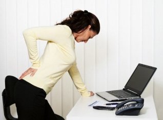
According to medical statistics, lower back pain in 80% of cases is caused by lumbar osteochondrosis. This occurs as a result of degenerative-dystrophic changes in this segment when the intervertebral discs and adjacent vertebrae are affected. Osteochondrosis of the lumbar spine (OBOP) manifests itself in various symptoms: pain of various natures, limited mobility, impaired sensitivity of the lower body and others. In the long absence of treatment, the degenerative processes spread to the vertebrae, reducing the ability to work, after which the patient may become disabled.
To avoid dangerous complications of lumbar osteochondrosis (LP), you should start a comprehensive treatment in 1-2 stages of the pathology. In advanced cases, when there are already irreversible changes in the disc or vertebrae, surgery is performed. To avoid osteochondrosis of the lower back and related complications, it is necessary to carry out its prevention.
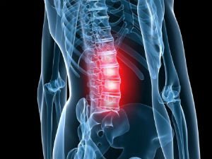
Development of lumbar osteochondrosis
To understand what is osteochondrosis of the lumbar spine, you need to study the structure of the spine. It consists of vertebrae between which cartilaginous pads (intervertebral disc) are placed. The disc is covered with a hard fibrous membrane (annulus fibrosus), inside which is the nucleus pulposus. This structure has a cushioning function and makes the spine more flexible.
Help. The lumbar segment of the spine is subjected to enormous stress on a daily basis, as it can support the weight of the upper body. Therefore, osteochondrosis of the lower spine is diagnosed more often than cervical, thoracic.
With regular loading of the spine, the discs shrink, lose a lot of fluid, their height decreases and the distance between the vertebrae decreases. The cartilage sheath becomes brittle, microcracks appear on its surface, through which the pulp nucleus protrudes over time. Upon further compression of the intervertebral discs, the outer shell ruptures and the gelatinous body falls off, forming a hernia. Then there is pathological mobility of the vertebrae, the load on the adjacent segments of the spine increases.
A little later, bone growths (osteophytes) begin to form at the edges of the vertebral bodies. In this way, the body tries to stabilize the spine.
Doctors distinguish 4 stages of osteochondrosis of the lumbar spine:
- 1 degree - problems with the discs begin, the central part dehydrates, flattens, cracks appear on the outer shell. There is a deleted current.
- Grade 2 - the cartilage sags, the vertebrae approach each other, become more mobile, the muscles and ligaments around the spine sag. Pain appears.
- Grade 3 - protrusions, hernias and subluxations of the vertebrae are formed. The pain increases, mobility is limited, the sensitivity of the lower body is impaired. Grade 4 osteochondrosis is characterized by the appearance of osteophytes, which can damage the spinal nerves and adjacent vertebrae. There is constant pain, severe neurological disorders and other complications, the risk of disability increases.
The easiest way to treat chondrosis of the lower back (stage 1), but to identify the disease at this stage is very difficult. Grade 2 intervertebral osteochondrosis is treated with conservative techniques. In stages 3-4, surgery may be required.
Help. According to statistics, OBO is more common in patients after 30 years. There are frequent cases of pathology in people after 20 years. Approximately 80% of 60-year-old patients suffer from this disease.
Causes
To understand how to deal with PKOP with osteochondrosis (lumbosacral spine), you need to know the reasons for it:
- Regular static or dynamic loading of the lumbar segment. The risk group for the development of osteochondrosis includes office workers, professional athletes (weightlifting), movers, builders and others.
- Poor posture, prolonged inappropriate posture.
- Genetic predisposition, abnormalities in the formation of vertebral bodies. This category includes youth software - curvature of the spine caused by pathologies of the spine.
- Spinal injuries.
- Hormonal imbalance, metabolic disorders, diseases of the endocrine glands, which disrupt the metabolism in the lumbar segment.
- Age-related changes in the body provoke disc wear.
- Tuberculosis of the bones, osteomyelitis (purulent inflammation of the bone tissue), ankylosing spondylitis (inflammation of the vertebrae and joints), rheumatoid arthritis, etc.
The disease is often caused by several reasons.
In addition, there are factors that provoke the development of lumbar osteochondrosis:
- Overweight.
- Passive lifestyle, prolonged sitting.
- Regular consumption of unhealthy foods (fatty, fried foods, confectionery, semi-finished products, etc. ).
- Lack of fluid, dehydration.
- Congenital disorders of the structure of the spine, such as an additional vertebra.
- Wear uncomfortable heels regularly.
- The period of pregnancy then increases the load on the spine.
- Sudden refusal to train professional athletes or excessive sports in people who have previously led a passive lifestyle.
- Smoking, frequent and excessive drinking.
There are many more factors that can cause degenerative-dystrophic processes in the lumbar spine. For example, flat feet, frequent hypothermia of the back, frequent stress, sleep disorders and others.
Symptoms
The symptoms of osteochondrosis of the lumbar spine are varied, they depend on the stage of pathology and the location of the affected area.
Doctors distinguish between reflex and compression syndromes (complex of symptoms) in OBOR. The former occur when the receptors on the outer shell of the discs, ligaments, joint capsules are irritated, and the latter when the nerve bundles, blood vessels and spinal cord are compressed.
There are such reflex syndromes of lumbar osteochondrosis:
- Lumbago. Shooting pain in the lower back with sudden movement or exertion. At the slightest attempt to move, the pain syndrome intensifies, so that the patient freezes in one position. The muscles in the injured area are very tense, on palpation the painful sensations become more pronounced. These manifestations are associated with the movement of the pulpal nucleus in the outer shell.
- Lumbodynia. The excruciating pain develops over several hours or days. Discomfort increases with movement, change in body position. Weakens when a person assumes a horizontal position with a roller under the lower back. When lifting a straight leg in this position, the pain intensifies (Lassegh's symptom). The degree of muscle tension is lower than in lumbago. Lower back mobility is limited.
- Lumbosciatica. Painful sensations (acute or painful) spread from the lower back to the lower body. There is an increase in this sign during movements. The pain is relieved by resting on the back. The muscles in the affected area are tense, the pain syndrome becomes pronounced on palpation.
The symptoms of compression syndromes depend on which parts of the lumbar segment are damaged. The characteristic signs are associated with compression of the spinal nerves by hernias, osteophytes, displaced vertebrae. This condition is called radiculopathy, in which the pain increases with the slightest movement, the muscles of the lower back are tense, and mobility is limited.
Clinical manifestations of compression syndromes depending on the damaged vertebrae of the lumbar segment:
- L1 - L3 - pain and tingling in the lumbar region, front and inner thigh, the patient has difficulty bending / unfolding the leg at the knee.
- L4 - the pain syndrome extends to the front of the thigh, down to the knee (back). Sensitivity is impaired in the same area.
- L5 - painful sensations radiate to the buttocks, outer thigh, descend on the front of the lower leg to the inside of the foot and big toe. Numbness is felt in the same area, it is difficult for the patient to bend the big toe.
- S1 - the pain spreads from the lower back to the buttocks, outer and back of the thigh, descends to the outer part of the lower leg, foot. Numbness is felt in the same areas, the calf muscles are weakened, so it is difficult for the patient to stand on his toes.
There is a risk of damage to several nerve bundles simultaneously, for example L5, S1. If the hernia moves backwards, it can compress the spinal cord.
Compression of blood vessels in the lower back increases the likelihood of weakening of the muscles of the legs, tingling in the lower extremities, impaired control over the process of urination and defecation. In men with OBO, erections are impaired, and in women the main symptoms may be supplemented by inflammation of the ovaries or uterus.
Diagnostic measures
To diagnose OBO, the doctor examines the patient, palpates the patient to determine the condition of the muscles and identifies the curvature of the spine. It is important to tell your specialist in detail about your symptoms to make it easier to diagnose.
Instrumental tests will help detect intervertebral osteochondrosis:
- X-ray of the lower back (front and side projection).
- Computer and magnetic resonance imaging.
The X-ray allows you to assess the structure of the EPP. In order to detect abnormal mobility of the vertebrae, X-rays are made in positions of flexion and extension. This examination allows us to notice that the intervertebral fissure has narrowed, the vertebral bodies have shifted and osteophytes have appeared at their edges. However, this diagnostic method is considered obsolete.
Today, CT and MRI are increasingly used to detect degenerative-dystrophic changes in the spine. These extremely informative studies make it possible to assess the condition of the vertebrae, discs, intervertebral foramina and spinal cord. With their help, protrusions, the direction of the hernia, the degree of compression of the nerve bundles, the spinal cord and blood vessels are detected.
Treatment
DRUGS FOR LEMAL OSTEOCHONDROSIS

Treatment of EPP osteochondrosis lasts from 1-3 months to 1 year. The success of therapy depends on the patient himself, who must strictly follow the doctor's recommendations. With self-medication, the patient's condition usually worsens.
Treatment goals:
- Stop or alleviate software symptoms.
- Identify the cause of the disease, try to exclude it from life.
- Eliminate the inflammatory process.
- Restores blood circulation, metabolic processes in the lumbar spine.
- Try to improve the condition of the damaged cartilage mucosa, stop further degenerative changes.
Complex therapy is recommended to achieve such goals. It usually starts with medication:
- Muscle relaxants. They relax the muscles and relieve pain and inflammation.
- NSAIDs. They have anti-inflammatory, analgesic, antipyretic action.
- Antispasmodics. They help to stop the spasms of smooth muscles, relieve pain.
- Anesthetics. They are used in severe pain syndrome in the form of therapeutic blockade.
- Glucocorticosteroids. They also help deal with pain. However, these drugs are able to destroy bones, so they are taken for a short time and only after approval by a doctor.
- Soothing. They relieve neuromuscular tension, improve sleep.
- Vitamins (group B, E, C, A). Restores the condition of the affected nerves, relieves pain.
Careful. NSAIDs should not be taken for gastritis or stomach ulcers, as they further damage the lining of the gastrointestinal tract.
In case of exacerbation, the patient is given injections and after relieving the main symptoms, he takes medication orally.
In addition, external agents (gels, ointments, creams, rubbing) are used.
The question of what to do in case of chronic osteochondrosis of the lower back is quite relevant. If OBOP has become chronic, after relieving the main symptoms of the patient are prescribed chondroprotectors, drugs that restore blood circulation, drugs based on vitamin B. They help to restore innervation, normalize blood supply to the affected area and prevent further development of the pathology.
The treatment of chondrosis of the lumbar spine (stage 1) is carried out with the use of chondroprotectors, which slow down the development of degenerative processes, accelerate the regeneration of cartilage. In addition, the patient is prescribed vitamin and mineral complexes. This form of osteochondrosis is the easiest to treat.
OTHER CONSERVATIVE TECHNIQUES
In case of acute chronic disease (osteochondrosis) of 1 - 2 degrees, the following treatment procedures will help to stop its development:
- Ultrasound therapy relieves pain and inflammation and normalizes blood flow to the affected area.
- Detensor therapy is a safe traction of the spine due to the weight of your own body, after which muscle tone is normalized and mobility is improved.
- Magnetic therapy reduces pain and inflammation of the muscles around the spine.
- Reflexology (inserting needles into bioactive points on the body) speeds up blood circulation, relieves inflammation and swelling.
- Manual therapy (impact on the affected area with the hands of a doctor) and massage normalize muscle tone, reduce the compression of nerve bundles, improve the nutrition of the intervertebral discs and restore the structure of the spine.
- Electrophoresis allows the delivery of drug solutions through the skin to bone and cartilage tissues. Drazonvalization improves blood circulation, metabolic processes, reduces pain, restores skin sensitivity.
There are much more effective procedures that will help improve the patient's condition in 5-15 sessions. The main thing is to get approval from a doctor before performing them.
SOFTWARE TREATMENT AT HOME
If you are wondering if it is possible to treat OBO at home, consult your doctor. If the specialist has given permission, then start therapy, which usually consists of the following points:
- Diet. If lumbar osteochondrosis is caused by impaired blood flow or metabolism, exclude from the menu fatty, fried, spicy foods, eggs and more. Fill the menu with fresh vegetables, fruits, lean meat, fish, dairy products. Give up alcohol, tonic drinks (tea, coffee). Drink filtered water, compotes, herbal teas.
- To restore blood circulation, apply or apply rubbing and compresses.
- Sleep on an orthopedic mattress, low pillow. If you have a sedentary job, buy a chair with a back that will support your spine. Wear special corsets or belts from time to time.
- Exercises will help strengthen the muscular corset, will relieve some of the load from the diseased spine. The complex for each patient is compiled individually by a doctor or instructor.
- Self-massage the lumbar region. However, ask a specialist how to do it right.
- Use folk remedies in the form of rubbing, compresses, baths and more.
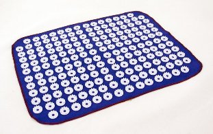
- The needle applicator is a plastic plate with many spikes, which improves blood circulation, metabolic processes in the damaged area, reduces muscle pain and relaxation.
And also at home you can use lotions with herbal decoctions, plasters.
Help. A novelty in the treatment of osteochondrosis is the massage bed, which is suitable for even the most disorganized patients.
Remember, however, that home treatment can only be done with your doctor's permission.
SURGICAL TREATMENT
Lumbar osteochondrosis surgery is prescribed if conservative techniques have proved ineffective for a long time. Also, surgery is indicated for involuntary urination, defecation and cauda equina syndrome (pinching of the nerves of the lower part of the spinal cord).
The following surgical methods are used to treat OBO:
- Spondylodesis - fusion of adjacent vertebrae.
- Feutextomy - removal of intervertebral joints that pinch the spinal nerve.
- Laminectomy is the removal of the lamina that covers the spinal canal, which compresses the spinal cord.
- Discectomy is the complete or partial removal of the intervertebral disc, which causes compression of the nerve root or spinal cord.
- Corpectomy - removal of the body of the spine and adjacent cartilage pads. Then the empty space is filled with a bone graft and 3 spinal segments merge.
Help. After the operation there is a risk of complications: spinal cord injury, nerve bundles, broken grafts, infections and more.
After treatment, you should undergo rehabilitation to speed up your recovery.
Complications
In the absence of appropriate therapy, the risk of such complications of lumbar osteochondrosis increases:
- Disc herniation, compressed nerve root or spinal cord.
- Prolonged inflammation increases the likelihood of developing radiculitis (inflammation of the nerve roots).
- Sciatica (inflammation of the sciatic nerve) in which there is severe pain and tingling in the lower limb.
- In case of impaired blood circulation in the spinal cord, the likelihood of compression myelopathy increases (compression of the spinal cord from various formations: bone fragments, hernia, tumors, hematoma).
- Cauda equina syndrome - compression of the roots of the lower part of the spinal cord, which leads to impaired function of the intestines, pelvic organs and lower limbs.
To avoid such complications, you should start treatment as early as possible.
Prevention
To avoid lumbar osteochondrosis, follow these rules:
- Lead a moderately active lifestyle (walk more often, exercise regularly, sign up for a pool).
- For sedentary work, warm up every 1. 5 hours.
- Sleep on an orthopedic mattress.
- Avoid excessive physical activity, lift weights only from a squat, before that put a special belt in the lower back.
- Buy orthopedic shoes.
- Eat right, take vitamin and mineral complexes as prescribed by your doctor.
- Learn to relax.
- Try not to hypothermia.
- Treat diseases that can cause OBO in a timely manner.
- Give up bad habits.
By following these recommendations, you can avoid degenerative changes in your spine and improve your health.
Most important
If you notice symptoms of lumbar osteochondrosis, see a doctor immediately. Self-medication can worsen your condition and cause complications. Lumbar chondrosis (stage 1) is treated with exercise, physiotherapy and chondroprotectors. At later stages, medications, massage, manual therapy, etc. are used. In the absence of positive dynamics for a long time or the appearance of neurological symptoms, the doctor may prescribe surgery. The patient should strictly follow the doctor's recommendations to speed recovery.